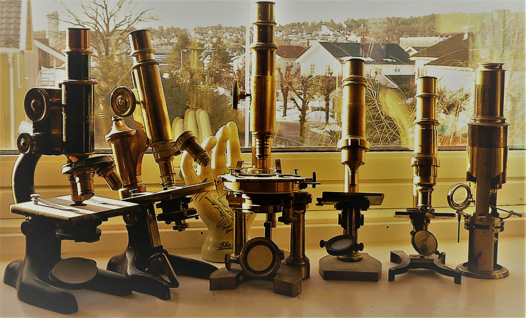
Mikroskopisk Microscopic
HISTOLOGY LABORATORY MANUAL
Vagelos College of Physicians & Surgeons
Columbia University
Patrice F Spitalnik MD
Histology Director
pfs2101@Columbia.edu
https://histologylab.ctl.columbia.edu/HistologyLabManual.pdf
Histology Guide. virtual microscopy laboratory
Atlas of Human Histology.
A Guide to Microscopic Structure of
Cells, Tissues and Organs. Robert L. Sorenson
http://www.histologyguide.com/about-us/sorenson-atlas-of-human-histology-chapters-1-and-14.pdf
Color Atlas of Cytology, Histology, and Microscopic Anatomy. Wolfgang Kuehnel, 2003 Thieme
Department of oncology Crimean State Medical University. Aliev K. A. Forskjellige organer – kreft
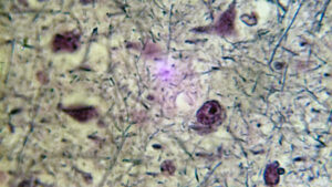 Hjerne
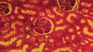 Nyre
Nyre
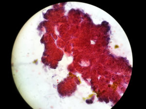
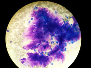 Pleomorft adenom i spyttkjertel (Cytologi)
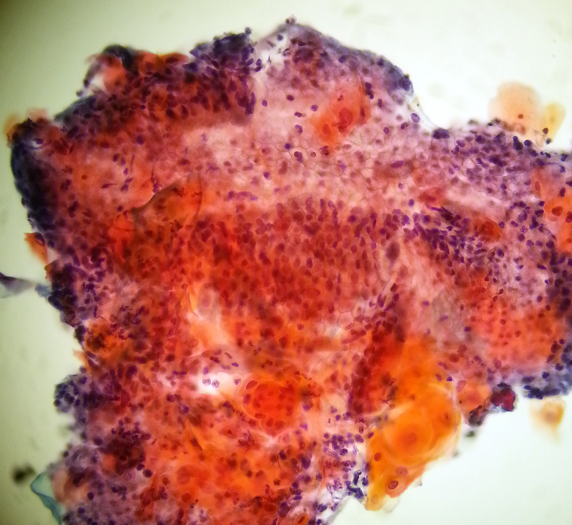
 Squamous cell carcinoma (Lung)
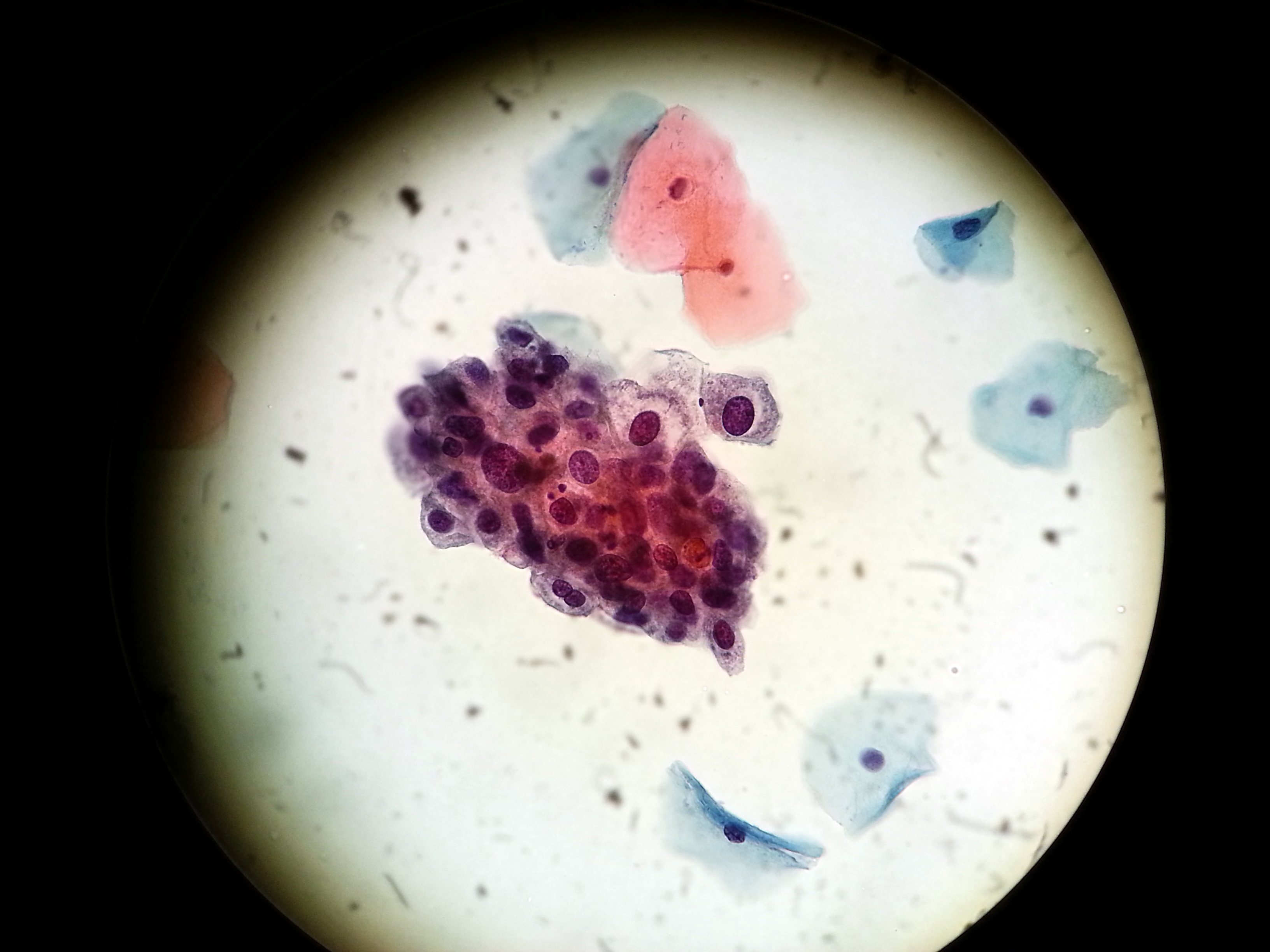
 Moderat atypi i gallegang (Cytologi)
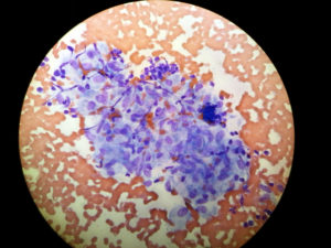 Sarcoidose, lunge (Cytologi)
 |
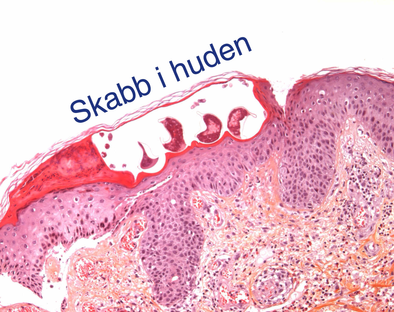
Skabb Mikroskopisk ned i huden

Rotifers under the microscope. Rotifers are microscopic animals found in aquatic environments all around the world. YouTube